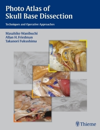
Photo Atlas of Skull Base Dissection
17
%
5269 Kč 6 383 Kč
Sleva až 70% u třetiny knih
With the many different approaches and dissection techniques to learn and practice, skull base surgery is one of the most difficult procedures to master. This photo atlas is an invaluable tool for understanding the complex anatomy and dissection process involved in skull base surgery. It offers a truly unique perspective in the presentation of step-by-step anatomic dissections as seen from the eye of an operating skull base surgeon. Logically organized and easy to use, this book offers concise coverage of different surgical approaches to the anterior skull base, anterolateral skull base, lateral skull base, posterolateral skull base, and posterior skull base. The book contains more than 1150 beautiful color photographs, as well as clinical information to aid in everyday practice.
Ideal for procedure preparation or reference, practitioners and residents in skull base surgery, neurosurgery, and head & neck surgery, alike will turn to this outstanding surgical guide time and again. A richly illustrated, step-by-step guide to the full range of approaches in skull base surgery, this book is designed to enable the surgeon to gain not only the technical expertise for common procedures, but to be able to confidently modify standard approaches when necessary. Full-color images of cadavers orient the surgeon to the clinical setting by presenting in precise detail the perspective encountered in the operating room. The images demonstrate surgical anatomy and the relevant structures adjacent to the exposures. Special emphasis on the relationship between the operative corridor and the surrounding anatomy helps the surgeon develop a clear understanding of whether tissues adjacent to the dissection can be exposed without complications.
Features:
-More than 1,000 high-quality images demonstrate key concepts
-Brief lists of Key Steps guide the surgeon through each step of the dissection
-Concise text supplements each photograph, providing descriptions of technical maneuvers and clinical pearls
-Coverage of the latest innovative approaches enables surgeons to optimize clinical techniques
Through detailed coverage of surgical anatomy and relevant adjacent structures, this book enables clinicians to develop a solid understanding of the entire operative region as well as the limits and possibilities of each skull base approach. It is an indispensable reference for neurosurgeons, head and neck surgeons, and otolaryngologists, and residents in these specialties.
| Autor: | Wanibuchi, Masahiko |
| Nakladatel: | Thieme, Stuttgart |
| Rok vydání: | 2008 |
| Jazyk : | Angličtina |
| Vazba: | Hardback |
| Počet stran: | 430 |
Mohlo by se vám také líbit..
-
Abdominal Ultrasound: Step by Step
Block, Berthold
-
Examination of Peripheral Nerve Injuries
Russell, Stephen M.
-
Caries Management - Science and Clini...
Meyer-Lückel, Hendrik
-

Fetoneonatale Neurologie
Jorch, Gerhard
-

Atlas of Sectional Anatomy
Möller, Torsten B.
-
Neck Rejuvenation
Gentile, Richard D.
-
Clinical Breast Tomosynthesis
Salkowski, Lonie R.
-
Rhinoplasty - Cases and Techniques
Godin, Michael S.
-

Textbook of Diabetic Neuropathology
Gries, Friedrich A.
-
The Temporal Bone
Sanna, Mario
-
The Chest X-Ray
Hofer, Matthias
-

Pediatric Plastic and Reconstructive ...
Greene, Arin K.
-

Hearing Aids
Dillon, Harvey
-
Cochlear Implants
Waltzman, Susan B.
-

Chinese Herbal Medicine
Hammer, Leon I.
-
Neurovascular Anatomy in Intervention...
Krings, Timo


