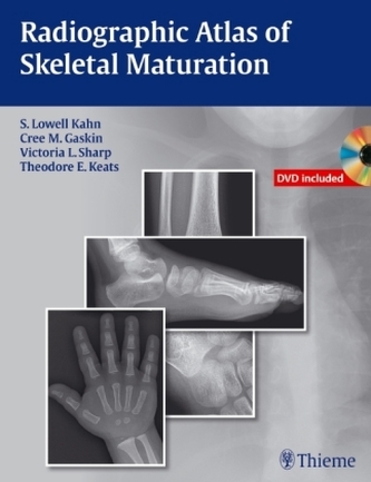
Radiographic Atlas of Skeletal Maturation, m. DVD-ROM
15
%
4876 Kč 5 745 Kč
Sleva až 70% u třetiny knih
ISBN 978-1-60406-571-8
Radiographic Atlas of Skeletal Maturation
S. Lowell Kahn Cree M. Gaskin Victoria L. Sharp Theodore E. Keats
DVD included
Nearly 2,300 images provide the reference standard for normal
skeletal maturation at every developmental stage
When dealing with the maturing skeleton and its many complex growth
alterations, physicians are constantly faced with the question, Is this image
normal? The Radiographic Atlas of Skeletal Maturation succinctly answers that
question by providing a comprehensive set of male and female reference images
for every age and body part. This allows physicians to quickly hone in on
normal ranges for the specific case they are reviewing, which is
particularly useful when called upon to read a pediatric skeletal radiograph in
the emergency room or while on call.
Special Features: - Nearly 2,300 high-quality images that provide instant
reference to normal views of the skeleton at every developmental
milestone-available in both the text and accompanying DVD - Multiple
projections at every age, sex, and body part combination so that the user can
match the reference points in the book to the case at hand and arrive at a solid
clinical interpretation (e.g., Is the small fragment of bone observed in a
7-year-old boy with an acute elbow injury a fracture or a normal developing
ossification center?) - Practical text layout organized by gender and body
part that provides quick access to images of normal development at any given
age - A software virtual skeletal survey demonstrates images of younger and
older individuals and crystallizes the subtle variations in growth patterns -
Powerful software package with advanced image enhancement tools allows
optimization of atlas image details for greater clarity. Compatible with
numerous image formats (including DICOM) allowing viewing and editing of outside
images - Convenient growth charts in the book and DVD for reference
This unique resource, with its vast collection of print and DVD images of
normal progressive skeletal development, gives physicians the full range of
comparative information they need to interpret pediatric skeletal radiographs in
any clinical setting. It is the reference standard for radiologists,
pediatricians, orthopedists, emergency room physicians, internists,
rehabilitation physicians, and training physicians who are called upon to review
a pediatric radiograph and confi dently make a diagnosis.
S. Lowell Kahn, MD, MBA, is Assistant Professor of Interventional Radiology
and Surgery, Tufts University School of Medicine, Baystate Vascular Services,
Baystate Medical Center, Springfi eld, Massachusetts Cree M. Gaskin, MD, is
Associate Professor of Radiology and Orthopaedic Surgery; Vice-Chair, Radiology
Informatics; and Director, Musculoskeletal Radiology Fellowship, University of
Virginia Health System, Charlottesville, Virginia Victoria L. Sharp, DO, is a
General Surgery Resident, Botsford Hospital, Farmington Hills,
Michigan Theodore E. Keats, MD (deceased), Former Alumni Professor of
Radiology, University of Virginia Health System, Charlottesville, Virginia
An award-winning international medical and scientifi c publisher, Thieme has
demonstrated its commitment to the highest standard of quality in the
state-of-the-art content and presentation of all of its products. Thieme's
trademark blue and silver covers have become synonymous with excellence in
publishing.
| Autor: | Kahn, S. Lowell |
| Nakladatel: | Thieme, Stuttgart |
| Rok vydání: | 2011 |
| Jazyk : | Angličtina |
| Vazba: | Hardback |
| Počet stran: | 608 |
Mohlo by se vám také líbit..
-
Normalbefunde in der Skelettreifung, ...
Kahn, S. Lowell
-
Abdominal Ultrasound: Step by Step
Block, Berthold
-
Head and Neck
Möller, Torsten B.
-

MRT der Wirbelsäule und des Spinalkanals
Forsting, Michael
-
Examination of Peripheral Nerve Injuries
Russell, Stephen M.
-
Caries Management - Science and Clini...
Meyer-Lückel, Hendrik
-

Neuroreha nach Schlaganfall
Mehrholz, Jan
-

In Führung gehen
Baller, Gaby
-

Altenpflege Lernkarten
Schön, Jasmin
-
Measurements and Classifications in M...
Waldt, Simone
-

Gehen verstehen
Götz-Neumann, Kirsten
-
Bestimmen von Sehhilfen
Methling, Dieter
-
Fetoneonatale Neurologie
Jorch, Gerhard
-

Aufteilung der endogenen Psychosen un...
Leonhard, Karl
-

Praxisnachweis Altenpflege
Baroud, Bianca
-

Atlas of Sectional Anatomy
Möller, Torsten B.


